


Coronavirus (COVID-19) Information and Resources
All of us at the UC Davis Eye center are here to serve our community. In these stressful times, the anxiety over the Corona virus pandemic and the necessary social distancing has affected all of us, both patients and providers. As always, our first concern is your welfare. For this reason and for your protection, we have reestablished clinical services, but only at 80% capacity in an effort to maintain proper social distancing throughout our clinical sites. We have undertaken an extensive cleaning protocol to ensure that each exam room is free of contamination. We are here for you, and look forward to the day when corona virus is a distant memory. Stay safe and stay well. Click here for more updates on COVID-19.
Mark Mannis, M.D.
Professor and Chair, UC Davis Eye Center
Leading the way in vision care and research
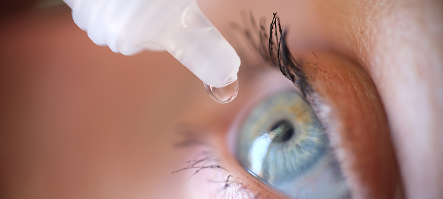
Welcome to the UC Davis Eye Center. Our vision is to be the world's transformational leader in collaborative vision research and in the development of cures for blinding eye disease from cornea to cortex.
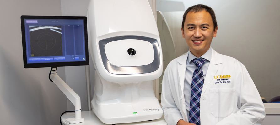
We will realize our vision through pioneering collaborative vision research, providing state-of-the-art, world-class eye care, and training superbly prepared ophthalmologists and vision scientists.
UC Davis Health Eye Center offers a full range of ophthalmic services for adults and children in Sacramento, Roseville, Folsom and Davis.
- UC Davis Eye Center
Ernest E. Tschannen Eye Institute
4860 Y Street, Sacramento, CA 95817
916-734-6602
Eye Center Optical Shop: 916-734-6300
Laser Eye Surgery (LASIK) Appointments: 916-734-6650
- UC Davis Eye Services in Davis
2035 Lyndell Terrace, Suite 100, Davis, CA 95616
916-734-6602 Appointments
530-757-6000 Office
Davis Optical Shop: 530-747-3360
- UC Davis Eye Services in Folsom
251 Turn Pike Dr., Suite 1070, Folsom, CA 95630
916-357-4880
Folsom Optical Shop: 916-357-4888
- UC Davis Eye Services in Roseville
1620 E. Roseville Pkwy, Suite 200, Roseville, Ca. 95661
916-771-0251 Appointments
Roseville Optical Shop: 916-746-6401
- UC Davis Student Health Services
Services Optometry Clinic and Optical Shop
For current UC Davis Students only
530-752-2349