
January 2024 – Presented by Dr. Heather Greene (Mentored by Dr. Chihong Heidi Zhou)
The patient is a 51-year-old male with a history of poorly controlled type 2 diabetes mellitus on insulin who presents to UC Davis Health with left parotid gland enlargement. He has noticed gradual enlargement of the area over the past year. He has pain at the angle of the mandible, which radiates to the neck. He noticed a slight decrease in size and alleviation of pain after a 2-week course of antibiotics.
Imaging of the parotid gland was performed. Representative sonographic and color Doppler sonographic images are shown in Figure 1 and Figure 2, respectively.
Following imaging, an ultrasound-guided fine needle aspiration biopsy was performed. Representative images of the resulting aspirate smears are shown in Figure 3.
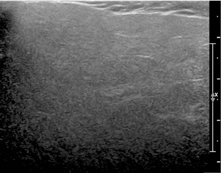 Figure 1: High-resolution sonographic image demonstrating diffuse parotid enlargement and hyperechogenicity without mass lesions, borrowed from Wen Y. and Goo HW., 2012.
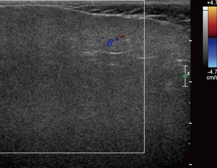 Figure 2: Color Doppler sonographic image demonstrating no evidence of increased blood flow, borrowed from Wen Y. and Goo HW., 2012.
Click on the images below to view larger.
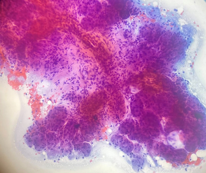
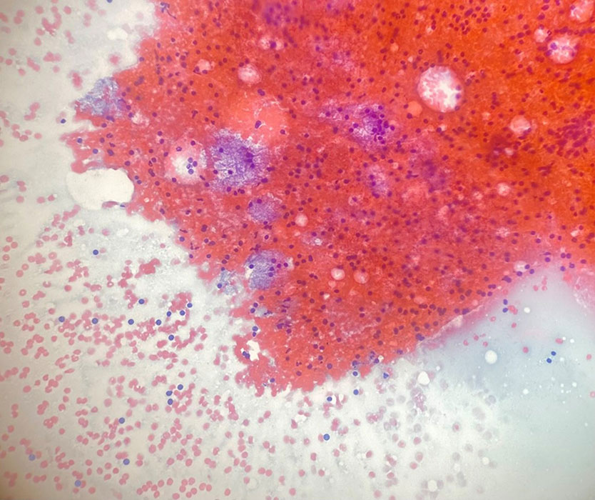
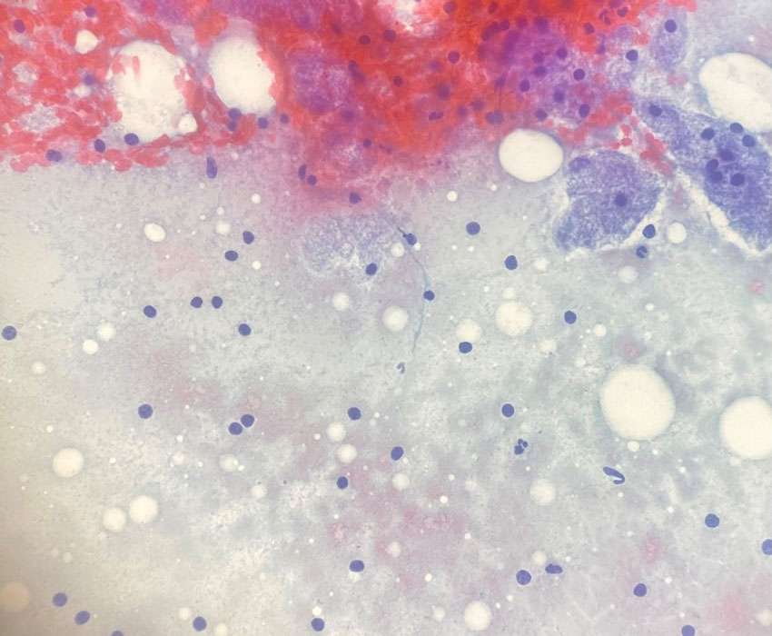
Figure 3: Aspirate smear, Papanicolaou (PAP) stain
 Meet our Residency Program Director
 LeShelle May
 Chancellor Gary May