
December 2023 – Presented by Dr. Lorene Chung (Mentored by Dr. Frank Melgoza)
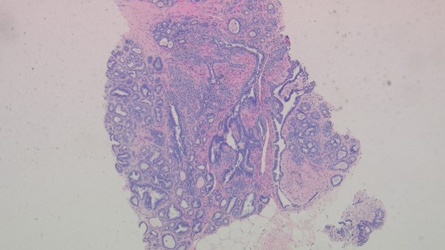
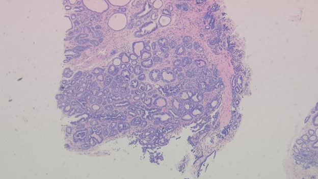
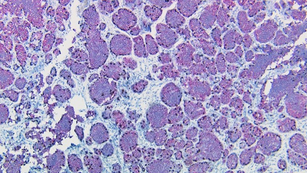
A 52-year-old, postmenopausal G1P1 female with a family history of breast carcinoma presents for routine mammogram and is found to have multiple left breast lesions. Menarche was at 14 years old. Imaging finds a 1.6 x 1.6 cm high density, partially circumscribed mass with microcalcifications located at 3:00, 6cm posterior to the nipple and a 1.4 cm mass at 12:00, 9cm posterior to the nipple line. Core needle biopsy is shown below. What is the most likely diagnosis?
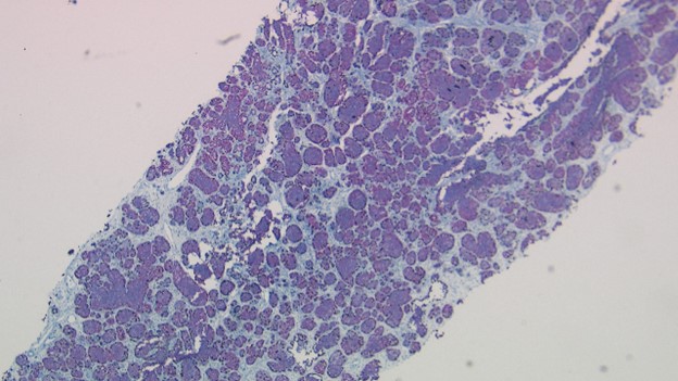
H&E
Click on the images to view larger.
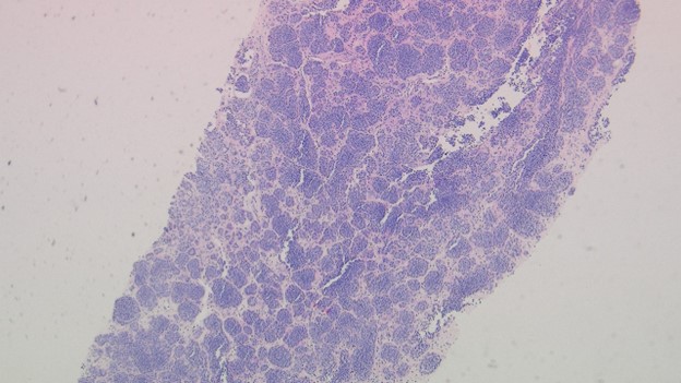
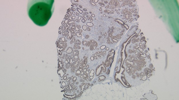
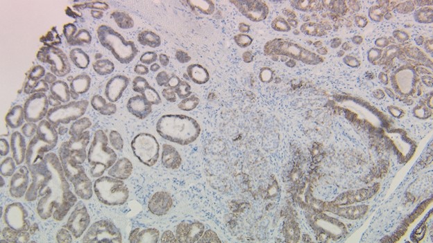
E-Cadherin
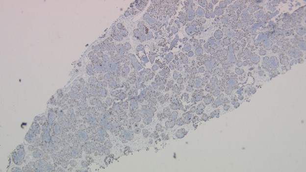
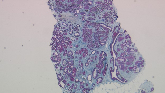
Tri-view Stain
Click on the images to view larger.
 Meet our Residency Program Director
 LeShelle May
 Chancellor Gary May