
November 2023 – Presented by Dr. Alex Chau (Mentored by Dr. Maaya Wilton)
A 77-year-old male with a history of COPD, AAA, and a 55-pack year smoking history was found to have a left lung lower lobe spiculated nodule on a routine low dose chest CT. He was referred for a CT guided biopsy (pictured below) which showed malignant cells. However, due to scant cellularity, IHC was unable to be performed. A PET scan was ordered and showed a hypermetabolic liver lesion measuring 3 cm. No other masses were present. The patient has no prior history of malignancy. The liver mass was biopsied and is shown below.
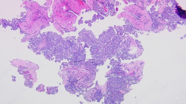 Liver, right lobe mass biopsy 40x
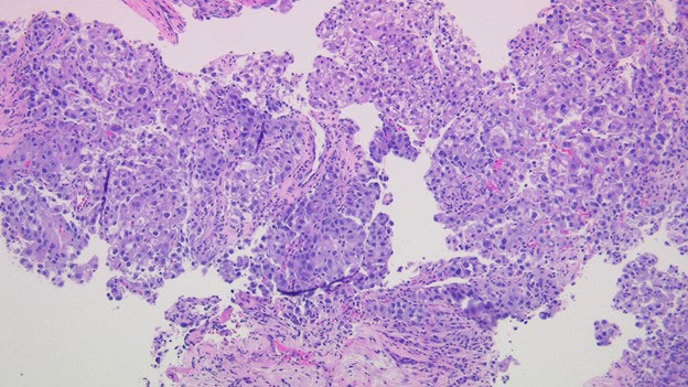 Liver, right lobe mass biopsy 100x
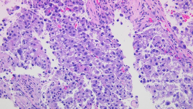 Liver, right lobe mass biopsy 200x
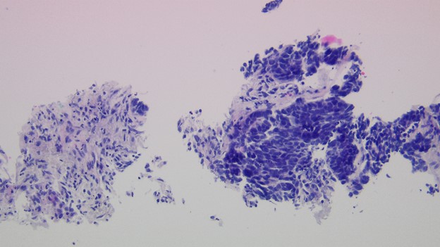 Previous lung biopsy 200x
 Meet our Residency Program Director
 LeShelle May
 Chancellor Gary May