
July 2023 – Presented by Dr. Jasper X. Zheng (Mentored by Drs. Bokhari and Tovar)
Q: The decedent was a 1 month old full term female infant who was born to a G3P3 female in her thirties by cesarean section. Other children were born healthy.
The decedent's Apgar scores were unremarkable with 8 and 8 at one and five minutes.
At birth, the decedent was noted with several anomalies: anorectal malformation, cloaca, rib anomalies, abdominal wall hemangioma, bilateral fetal choroid plexus cysts, and postnatal level 1 urinary tract dilation of left kidney.
The decedent experienced immediate post birth significant desaturation that led to the discovery of significant pulmonary hypertension. Medical intervention, including extracorporeal life support, was provided but was futile. As the decedent's condition deteriorated, the family elected to limit suffering by compassionate extubation. The decedent passed away peacefully.
Autopsy was performed and the lungs weight and grossly appear normal.
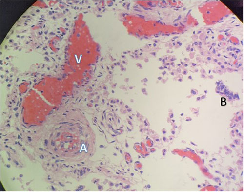
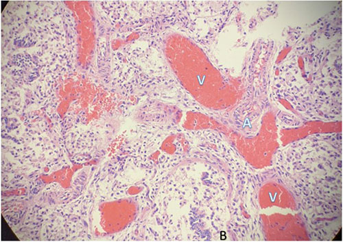
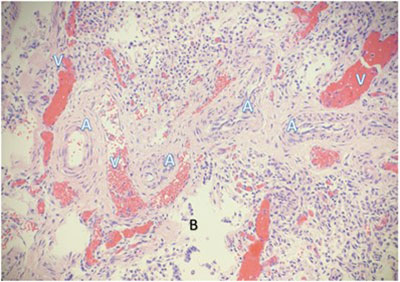
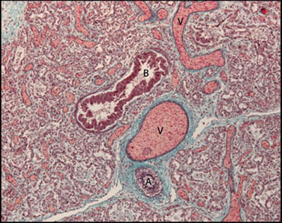
Histomorphological examination of the pulmonary system showed the following (Hematoxylin and Eosin at original magnification of 400x):
V = Pulmonary vein
A = Pulmonary arteriole
B = Bronchial epithelium
Immunostaining for CD31 highlighting the reduced number of alveolar capillary endothelial cells located away (arrows) from the inner side of the alveoli (dashed lines).
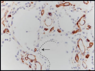
Source: Slot, E., et. al., doi: 10.1177/2045894018795143. PMID: 30058937.
Trichrome stained lung tissue
Source: Slot, E., et. al., doi: 10.1177/2045894018795143. PMID: 30058937.
 Meet our Residency Program Director
 LeShelle May
 Chancellor Gary May