
January 2022 – Presented by Dr. Luke Dang (Mentored by Dr. Kurt Schaberg)
Clinical Presentation
The patient is a 4 year-old male with a history of prematurity and cloacal exstrophy, status post multiple prior reconstructive surgeries, now presenting for urinary diversion with ileal conduit. Imaging is significant for one atrophic kidney, while the contralateral kidney is notable for hydronephrosis of the collecting system. A nephrectomy specimen (the atrophic kidney) is received by pathology and grossly is without mass lesions.
Microscopic
Figure 1. Disorganized architecture is present, kidney lacks usual collecting system (H&E, 2x)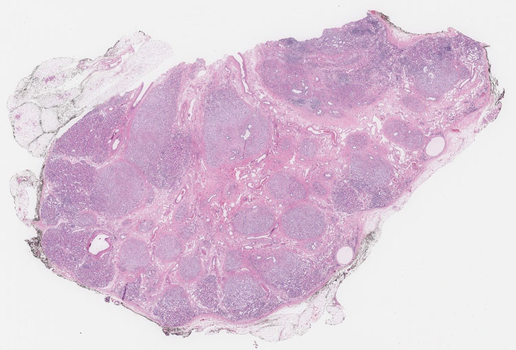
Figure 2. Immature tubules with surrounding collarette of spindle cell stroma (10x, 4x).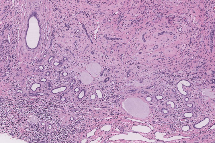
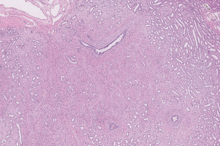
Figure 3. Focal islands of cartilage are present (H&E, 10x)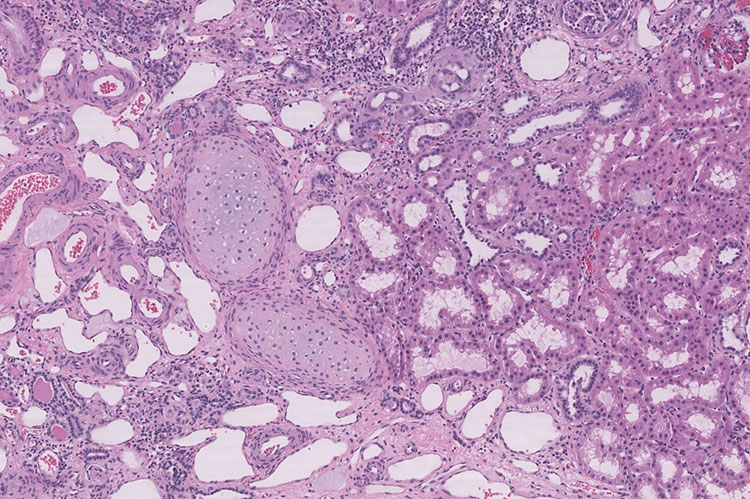
 Meet our Residency Program Director
 LeShelle May
 Chancellor Gary May