
March 2021 – Presented by Dr. Jake Donnelly (Mentored by Dr. Kurt Schaberg)
Clinical History
A 75-year-old male presented to the hospital with early satiety and melena. An upper endoscopy was performed and revealed a large proximal gastric cancer. Staging studies showed only regional disease and the patient was started on chemotherapy. Subsequently, the tumor was excised en bloc with a total gastrectomy.
Microscopy
Histologic sections of the stomach masses demonstrate invasive malignant cells arranged in small nests or fused glands with an associated prominent lymphocytic infiltrate and intraepithelial lymphocytes.
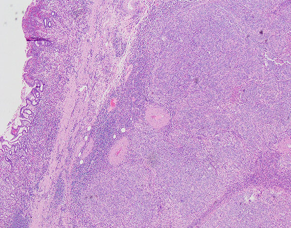
Figure 1. H&E 4x (Highlighting the tumor infiltrating lymphocytes)
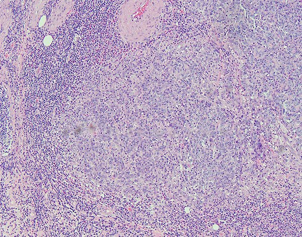
Figure 2. H&E 10x (Highlighting the tumor infiltrating lymphocytes)
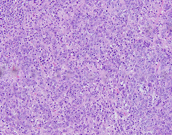
Figure 3. H&E 20x (Highlighting the tumor infiltrating lymphocytes)
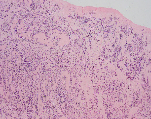
Figure 4. EBER ISH 2x
 Meet our Residency Program Director
 LeShelle May
 Chancellor Gary May