
Resident Program - Case of the Month
January 2019 - Presented by Dr. Miao Tian (Mentored by Dr. Dorina Gui)
Clinical History
A 72-year-old man presented to the emergency department with a chief complaint of abdominal pain, vomiting, poor appetite and weight loss. CT demonstrated thickening of the duodenum and a right adrenal mass. He underwent endoscopy with biopsy showing duodenal adenocarcinoma. For his adrenal mass, he was noted to have mildly elevated urine catecholamines and metanephrines. He also has elevated serum metanephrines. The patient denied hypertension, severe headache or palpations. He subsequently underwent pancreaticoduodenectomy and right adrenalectomy. The histopathology confirmed the duodenal adenocarcinoma. However, the adrenal mass showed unusual histological patterns.
Pathological Findings
Gross examination revealed a 4.0 x 3.5 x 2.7 cm ovoid, well circumscribed, firm, yellow mass arising from the medulla of the adrenal gland. No necrosis or hemorrhage is grossly identified.
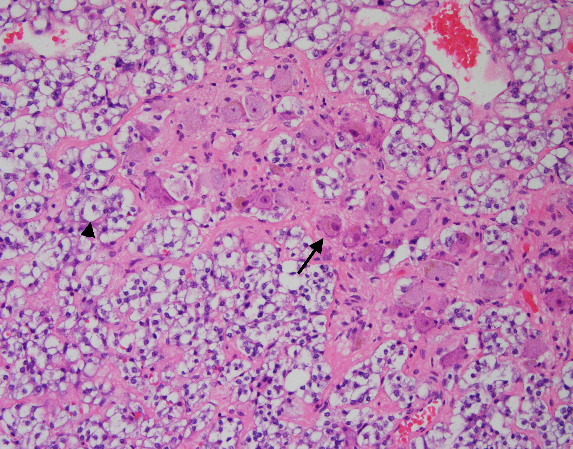
Microscopically, the specimen showed two irregularly distributed distinct tumor patterns (Figure 1). One pattern shows nests of tumor cells (Zellballen pattern). They have abundant cytoplasm and round to oval nuclei with inconspicuous nucleoli (Figure 1, arrowhead). The second pattern contains fibrillary stroma with spindle-shaped cells and clusters of large polyhedral tumor cells (Figure 1, arrow) with abundant eosinophilic cytoplasm, prominent nucleoli and occasional multinucleation.
Click on image to enlarge
| Figure 1 - H&E |
 |
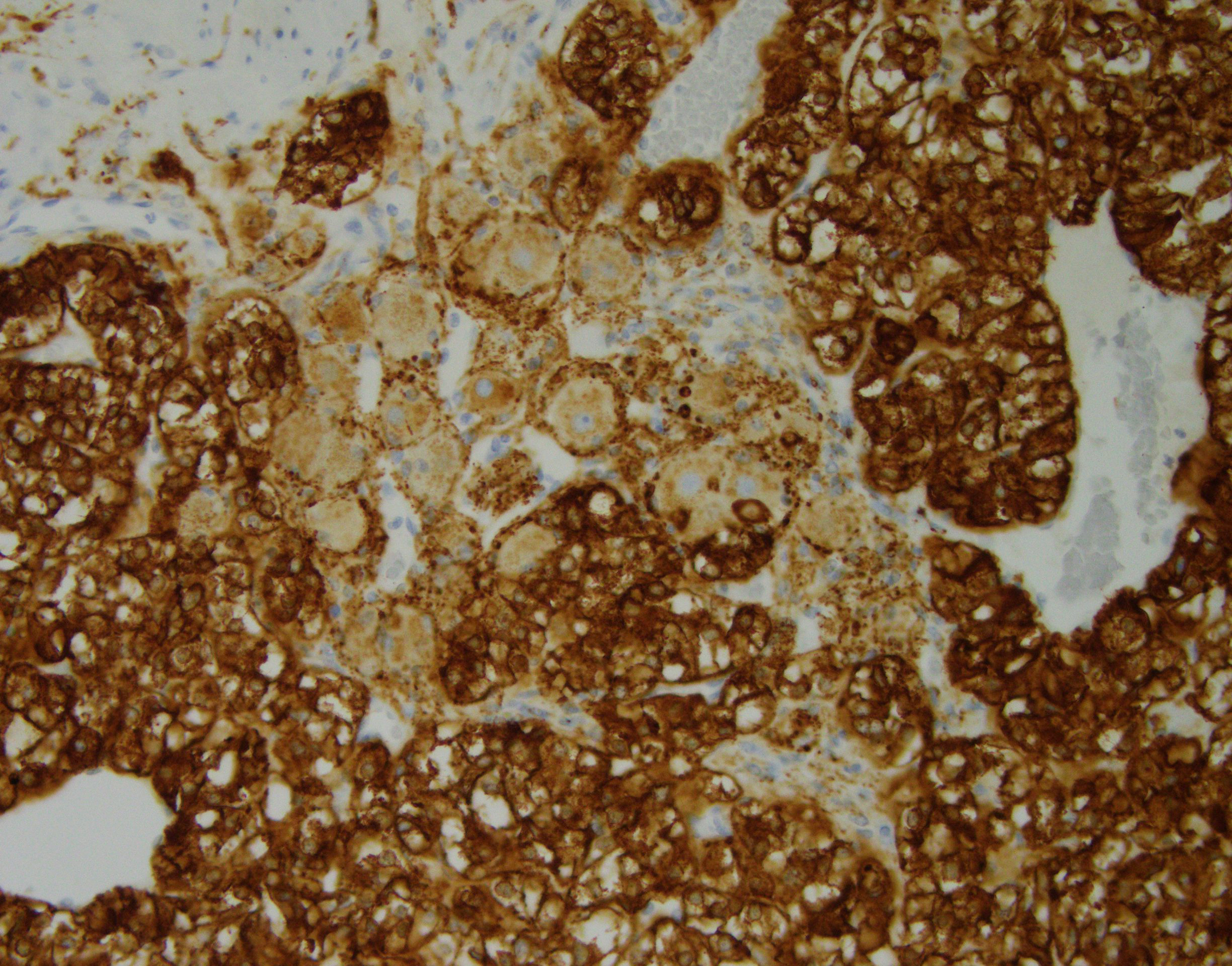
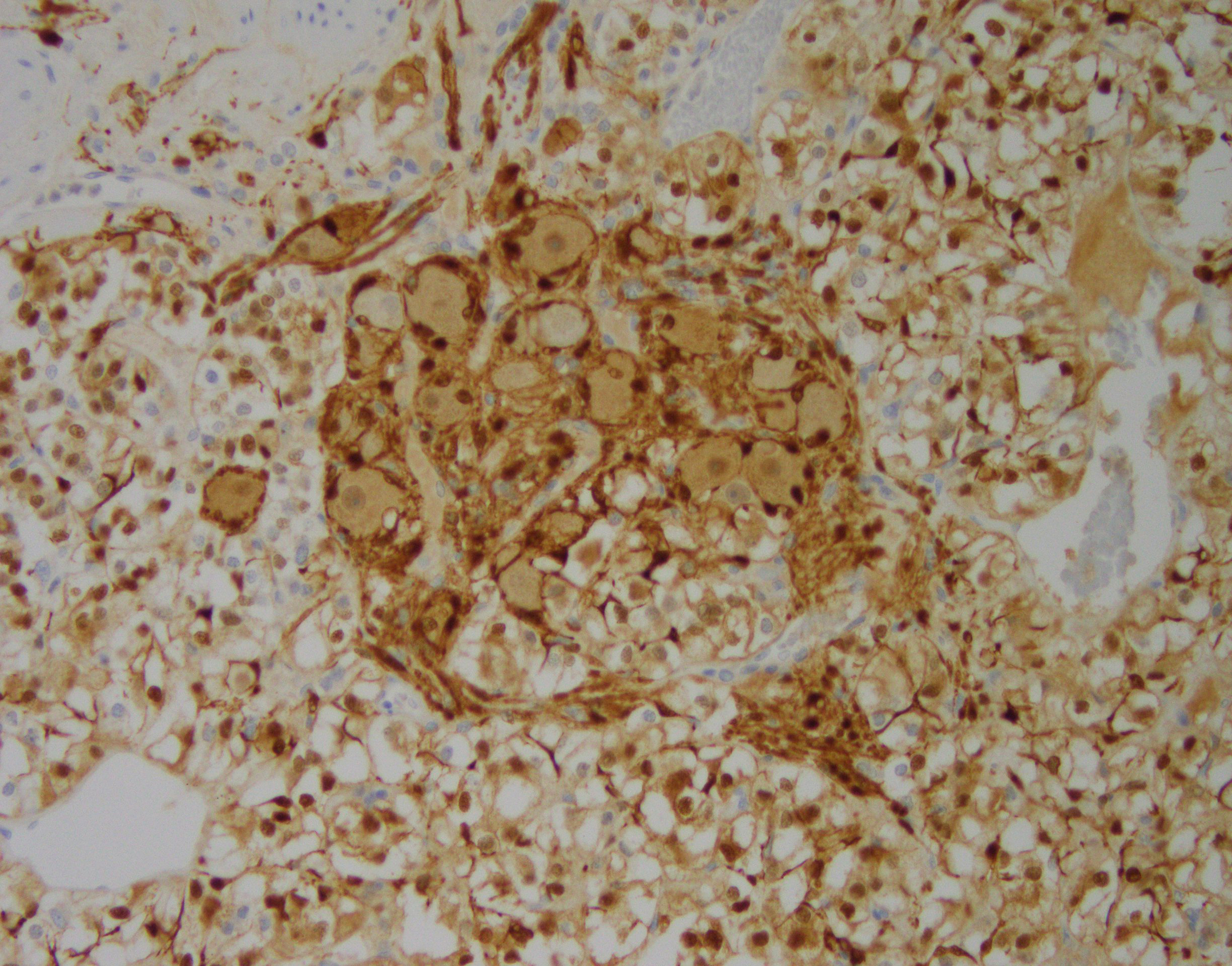
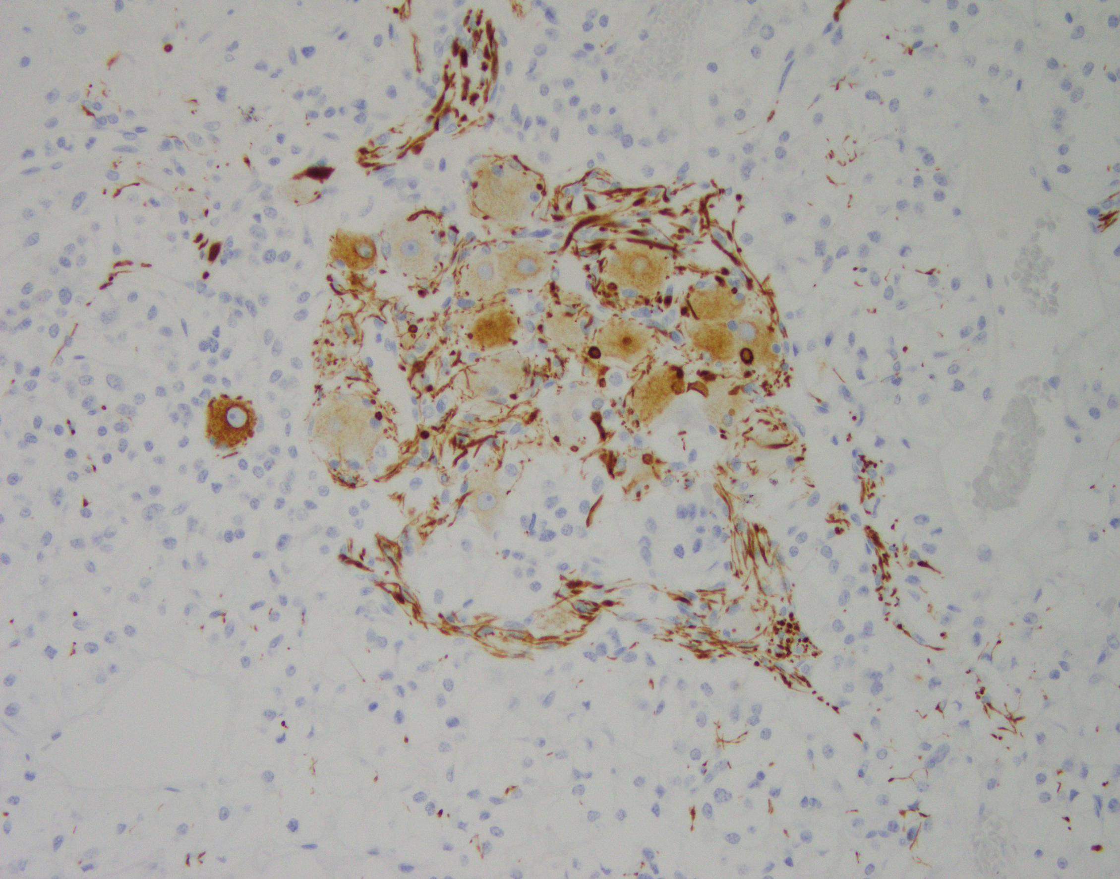
Synaptophysin immunohistochemical stain (Figure 2) reveals the different components of the composite tumor: intense cytoplasmic staining in the pheochromocytoma cells and weak staining in the neuronal cell bodies. S100 immunostain (Figure 3) highlights the zellballen pattern typical for pheochromocytoma and sustentacular cells. Immunostaining for the neurofilament protein (Figure 4) highlights axon-like processes. Cell bodies of both pheochromocytoma cells and neurons show weak or absent neurofilament staining.
Click on image to enlarge.
| Figure 2 Synaptophysin |
Figure 3 S-100 |
|
 |
 |
|
|
Figure 4 |
What is most likely the diagnosis for his adrenal mass?
Choose one answer and submit.
 Meet our Residency Program Director
 LeShelle May
 Chancellor Gary May