
Examples of Dr. deVere White's Research
RO1CA136597-01 National Cancer Institute: "Identification and characterization of the functional role of miRNA in prostate cancer".
Our hypothesis is that the expression of miR-125b in CR CaP cells is controlled by aberrantly-activated AR, that miR-125b facilitates CR growth of CaP cells by repressing the function of the p53 network, and that miR-125b has potential as a biomarker and a therapeutic target for the management of patients with CaP.
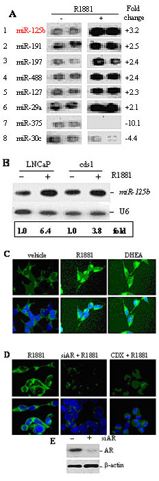
Androgen upregulates miR-125b.
A) Differentially-expressed miRNAs in LNCaP treated with 5.0 nM R1881 or ethanol vehicle.
B) Northern analysis of miR-125b in LNCaP and cds1cells treated with 5.0 nM R1881 or vehicle. U6 RNA is loading control.
C) The level of miR-125b were detected using a Fitc-LNA-miR-125b in LNCaP cells treated with ethanol vehicle, R1881 (5.0 nM) or DHEA (1.0 µM).
D) The level of miR-125b was detected in siAR- or CDX-treated LNCaP cells in the presence of 5.0 nM R1881. In both C & D, the fluorescent signals (green) are cytoplasmic in location. The nuclei were stained using dapi (blue). The experiment was performed twice with similar results each time.
E) Western blot analysis of the AR knockdown using siAR.
PC074103 Department of Defense-Prostate Cancer Research Program
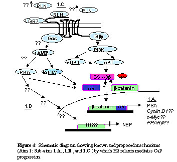
Functional validation of H2 relaxin, and its downstream effectors, as mediators, therapeutic targets and potential biomarkers of prostate cancer progression". Understanding the mechanism(s) by which H2 relaxin facilitates AI CaP could potentially allow for the design of effective therapies to treat this at present incurable disease and provide a new biomarker for CaP progression. The ability to predict the progression to AI CaP would be invaluable in determining appropriate patient treatment options.
PC080488 Department of Defense-Prostate Cancer Research Program
Deregulation of miRNAs contributes to development and progression of prostate cancer. The major goals of this project are to elucidate the mechanism by which miR-125b contributes to pathogenesis of CaP, evaluate the effects of miR-125b on prostatic tumorigenesis and AI growth, and to determine the application of miR-125b as a biomarker for CaP.
 |
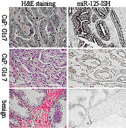 |
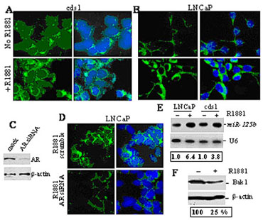
| Fig. 4. A,B&D) LNA-ISH analyses of miR-125b in cytoplasma (green). The nuclei were stained using dapi (blue); C) Western blotting; E) Northern blot analyses; F) Western blot analysis. |