

Tumor on a chip:
a new tool to personalize cancer care
Researchers and clinicians are learning how to personalize cancer treatments by adopting next-generation sequencing (NGS), mathematical models and other approaches to ensure that each patient gets the ideal combination of therapies.
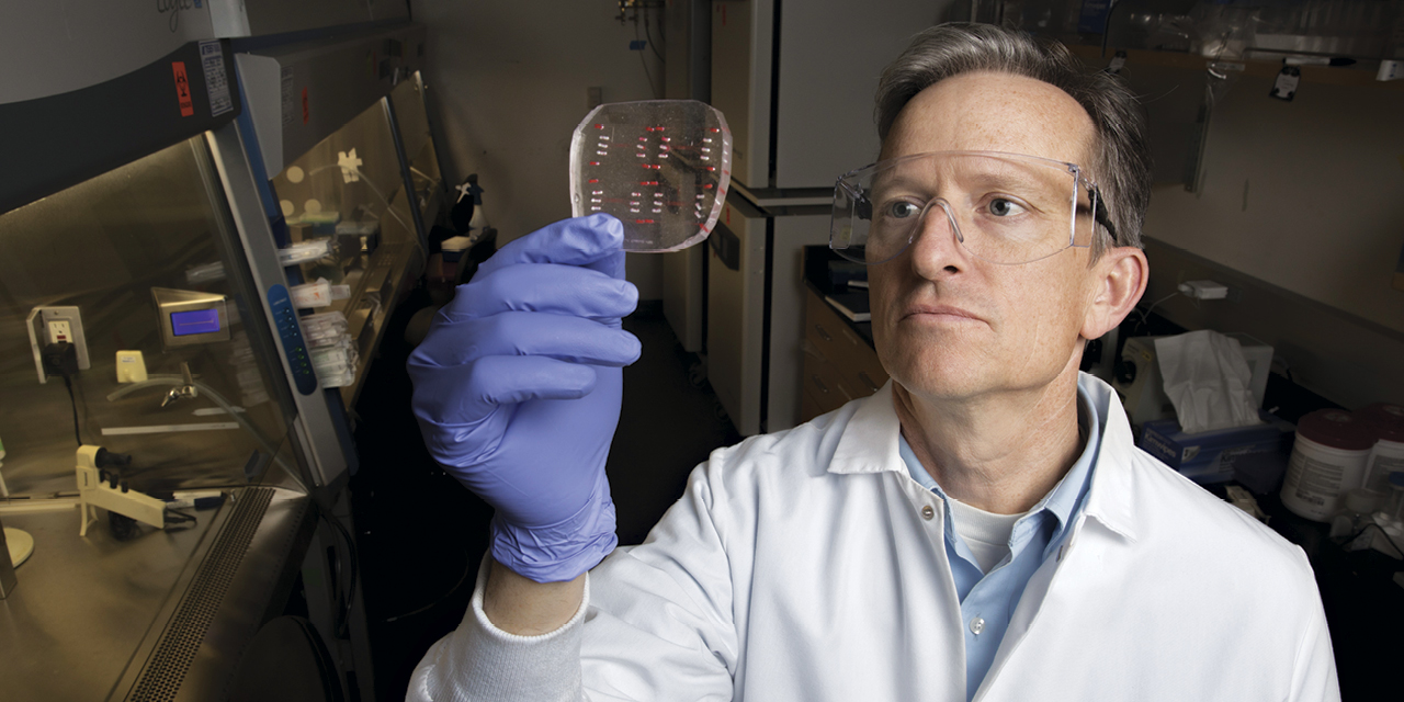
UC Davis biomedical engineer Steven George has a different strategy: create microtumors from patient samples and test drugs against them.
“We can put primary human cancer from a patient’s biopsy in a small, microfluidic device with perfused human capillaries and deliver drugs through the capillaries,” said George, professor in the Department of Biomedical Engineering. “It would give us an idea if the cancer is sensitive or resistant to a certain drug even before it is given to the patient.”
The device could help address a fundamental challenge in oncology: Cancer is not one disease but many. Consider the variations among breast cancers, such as ductal carcinoma in situ, invasive ductal carcinoma and triple-negative tumors. Each type might require a different therapeutic approach, and the complexity only increases. Cancers are chaotic, and even similar tumors often have different mutations, challenging physicians to find the most optimal therapies for each patient’s unique cancer.
NGS has been enormously helpful. By identifying specific mutations in tumors, clinicians can assign treatments that target those variations. But NGS has its own shortcomings. Sometimes targeted therapies fail, even when the tumor sequence indicates they should work.
George believes his technology could augment NGS and other systems to provide real-time feedback on which agents work for a particular tumor.
Drugs vs. tumors
George and colleagues have spent more than a decade developing this tumor-on-a-chip technology. The device, made from a flexible polymer — like a firm rubber — can fit into the palm of your hand. Microfluidic channels, about the width of a human hair, are connected to engineered human capillaries. Together, the channels deliver tumor material, the extracellular matrix and nutrients that support the tumor.
The capillaries are a key refinement, helping these 3D micro-tumors better replicate conditions inside the body. The tumor specimen itself is tiny, smaller than a cubic millimeter, making it incredibly helpful for clinicians, since acquiring large amounts of biopsied tumor tissue can be difficult. In addition, the platform’s small size will help researchers test multiple agents on different samples simultaneously.
“We can put tissue into the device and test several different drugs,” says George. “We could potentially screen a panel of drugs or test the same one at different concentrations. There are a number of choices in the neoadjuvant (prior to surgery) setting for breast cancer. If the tumor is more sensitive to one agent over another, then you would choose that drug.”
Immunotherapy
In addition to testing chemo or targeted therapies, the device could also help determine whether immune therapies can help specific patients. The first generation of these drugs, called checkpoint inhibitors, has been both miraculous and disappointing. For the 20–25 percent of patients who respond to these treatments, tumors evaporate. The rest see little to no effect, and the side effects can be significant.
Having the ability to test checkpoint inhibitors, or other immune system-targeting drugs, on live tumor tissue could help determine if a patient will respond.
This could be both a short- and long-term approach. George’s lab is currently modeling different ways to use stem cells to test immunotherapies, using cells from the patient to create induced pluripotent stem cells (iPSCs), which can then be coaxed in culture to form essentially any tissue. Because these cells come from the patient, and not a donor, they could provide more precise information on how that patient’s immune system will react to therapy.
“You would have a genetically matched in vitro system to test immunotherapy or other agents,” says George.
This is not a fast process — it would take three to six months to transform a patient’s cells into the target tissue. However, this method could give physicians a head start should the patient’s cancer return, which it often does.
“If we can give the clinician useful information six months later, they can know what to do if the cancer comes back,” says George.
Drug discovery
Looking down the road, a tumor on a chip could be a great advantage for drug discovery. While animal models can be very useful, they don’t quite replicate humans. As a result, many drugs that work in animal models fail in human clinical trials.
“The success rate is low because predicting drug efficacy can be quite difficult,” says George. “With our platform, you could have better information to choose which drugs go into clinical trials. So far, pharmaceutical companies have been interested in using it to test lead compounds prior to clinical trials.”
George and colleagues are still refining the system in the lab, and he has co-founded a biotech company, Kino Biosciences, to help commercialize the technology. And while the device has obvious applications in cancer, there are other potential applications.
“Eventually, our technology could be useful to test drugs for other diseases that involve the immune system,” says George. “We are currently exploring rheumatologic diseases, organ transplantation, multiple sclerosis and type I diabetes.”