|
WHEN WORLDS COLLIDE
Researcher and breast cancer survivor improves cancer imaging.
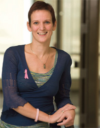
When Julie Sutcliffe was diagnosed with breast cancer three years ago, she found herself the beneficiary of her own experiments and the expertise of her colleagues.
As a researcher, Sutcliffe was testing novel cancer drugs and creating new compounds that would facilitate clearer imaging of cancer cells.
As a patient, she enrolled in a clinical trial of a new breast CT scanner – custom built by UC Davis biomedical engineering Professor John Boone – that provides comfortable, accurate imaging of breast tumors. Another colleague and now great friend, Richard Bold, was the surgeon who removed her tumor. As part of her treatment plan, she was given the drug Herceptin, which helps prevent breast cancer recurrence. Sutcliffe was previously involved in the preclinical testing of this compound.
"It was quite a balancing act to be a patient, a colleague and a scientist all at the same time," says Sutcliffe, an associate professor in the Department of Biomedical Engineering and director of the cyclotron and radiochemistry facility for the Center for Molecular and Genomic Imaging. "It gave me a whole new perspective," she says of her battle with the disease.
With her cancer now in remission, Sutcliffe is driven more than ever by her passion for improving molecular imaging for cancer detection and treatment planning. She is working to develop new short-lived radioactive imaging agents for use in PET (positron emission tomography) scans.
PET is a powerful imaging tool that allows physicians to precisely pinpoint tumors and see them in action, providing valuable information to help fight the disease. Using this technology requires selective imaging agents – radioactive compounds called "radiotracers" – that provide very high contrast between diseased and healthy tissue and the means to examine the biology of cancer.
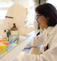
Sutcliffe's laboratory focuses on finding new selective agents for cancer imaging that rapidly distribute throughout the body and clear quickly from nontarget tissues. The specific agents Sutcliffe focuses on bind to certain molecules on cancer cells, literally making the cancer glow on a PET scan's computer-generated, three-dimensional images.
Currently, there is only one FDA-approved PET radiotracer used to image and stage cancers. This compound mimics glucose and, therefore, goes to any cell that metabolizes glucose. Because it is metabolized by cancer cells at about 25 times the rate of normal cells, glucose is excellent for detecting primary cancers and metastases. The problem is that this agent cannot distinguish disease from infection.
"PET scans of glucose metabolism are highly informative, but there is a need to image more specific biological processes of cancer with more selective targeted imaging agents," Sutcliffe says. "Right now, we're way behind on the translation of these compounds from the bench to the bedside to realize the full potential of PET."
From chemistry to patients
Sutcliffe and her lab team have already developed one new promising imaging agent that binds to a particular protein found almost exclusively on the surface of cancer cells. The protein – alpha-v-beta-6 – belongs to a family of proteins called integrins found on the surface of many different kinds of cancer cells, including breast, pancreatic, colon and lung cancer. Its presence is considered a reliable predictor of poor outcome.
 |
 |
| |
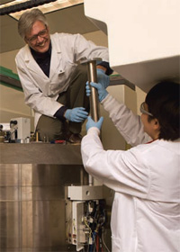
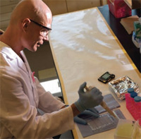
Postdoctoral researcher Sven Hausner is designing imaging probes to target diseases such as head and neck cancers, pancreatic cancer and breast cancer. |
|
|
"Knowing that cancer is present and the pathological course it is likely to take are powerful tools in developing successful treatment plans," says Sutcliffe. "Our work brings much greater precision to diagnosis and treatment of the disease."
The imaging agent Sutcliffe designed uses part of a protein made by a virus – the one that causes foot-and-mouth disease – that selectively binds to the alpha-v-beta-6 integrin. She radio-labeled this peptide using 18F fluoride and, in 2007, showed for the first time that it was highly visible in tumors using a small-animal PET scanner developed by colleague Simon Cherry, director of the UC Davis Center for Molecular and Genomic Imaging.
Based partly on her integrin results, Sutcliffe received a highly competitive grant from the U.S. Department of Energy to produce more high-quality imaging agents for use in humans. According to Sutcliffe, the agents she's developing could be used to image any cell surface receptor associated with a particular disease. To that end, she has received a UC Davis Cancer Center Support Grant to develop targeted molecular imaging agents for a marker of tumor angiogenesis – the process by which tumors form blood vessels. A marker for these tumor-associated blood vessels would allow physicians to see how a tumor is able to sustain itself and, potentially, allow them to track the efficacy of anti-angiogenic drugs intended to "starve" the tumor by shutting off its blood supply.
"Julie is driven by her passion and will to win against all odds," says Michael Phelps, Norton Simon Professor and chair of Molecular and Medical Pharmacology at UCLA. "These personal qualities, combined with her scholarship, make her a remarkable scientist. Julie has developed truly innovative approaches to rapidly develop diverse arrays of imaging probes for PET, providing the means to examine diverse arrays of therapeutic targets in cancer patients. Her unique approach expands the value PET molecular imaging diagnostics can provide to better manage the biology of cancer to the benefit of cancer patients. All of this is part of the wonderful evolution of Julie as a person and as a scientist. It is my privilege to be her colleague and her friend."
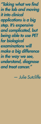
Sutcliffe expects to begin human clinical trials of the integrin-targeting imaging agent within the next 18 months. Grants from the National Institutes of Health will help fund a new radiochemistry laboratory and provide an ultra-clean environment to derive cellular products for patients involved in clinical trials. "Taking what we find in the lab and moving it into clinical applications is a big step," Sutcliffe explains. "It's expensive and complicated, but being able to use PET for biological examinations will make a big difference in the way we see, understand, diagnose and treat cancer."
Back to the clinic
Getting back to clinical work has been Sutcliffe's goal since arriving in 2002 at UC Davis. Her path to academia was quite unconventional. At age 22 and with just an undergraduate degree in chemistry, she joined the world-renowned PET group at the Hammersmith Hospital in London. She spent two invaluable years there gaining a wealth of experience and skills at an emerging time for clinical PET. She was fascinated with the radiochemistry and by the almost science fiction-like quality of the imaging machines used on patients.
"This was a brand new field and few people knew how to do the medicinal chemistry I was learning. These were fun, exciting times," Sutcliffe says. Two years later, she applied for and got a job running the radiochemistry lab at the prestigious St. Thomas' Hospital in London
Eventually, Sutcliffe realized that becoming an innovator in her field meant getting more degrees. She went to night school, earning a master's degree in synthetic organic chemistry. Later, she enrolled at King's College School of Pharmacy, conducting her Ph.D. research in her own lab.
That was when Cherry asked Sutcliffe to come to UC Davis, and she jumped at the chance to work with a recognized leader in imaging science. She filed her dissertation and, a month later, arrived at UC Davis where her work has so far focused on bench science and pre-clinical trials.
"I love my work, but I must admit that I have missed the clinic," Sutcliffe says. She is excited that next she will see how her imaging agents perform in patients. "Now I can do what I really came here to do, which is show how the work in the lab connects with improvements in cancer care."
|