Residency Program - Case of the Month
July 2013 - Presented by Mahan Matin, M.D.
Clinical history:
A 3-year-old boy presented with an enlarging mass in front of his ear. There was no bleeding or bruising, change in visual acuity, fever, pain, or drainage from the ear. He had a history of RSV infection and asthma 1 year ago. On physical exam the physician noticed lymphadenopathy in level 2 on the right side of the neck and also posterior cervical chain in level 5. The cranial nerves II – XII were intact.
Neck CT scan with contrast showed a 3.8 x 2.2 cm enhancing mass centered in the R temporal bone, extending into the middle ear and mastoid air cells (radiologic images). The radiologic differential included eosinophilic granuloma, lymphoma, and metastatic neuroblastoma.
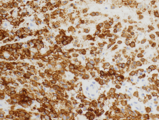
Fine needle aspiration was performed on the temporal mass. The histology is shown below in images 1-3. A few months later a biopsy was taken from the mass. The histology is shown below in images 4-8.
Microscopic Photographs:
| Image 1 | Image 2 | Image 3 |
 |
 |
| Image 4 | Image 5 | Image 6 |
 |
 |
 |
| Image 7 - s100 | Image 8 - CD1a | |
 |
 |


 Meet our Residency Program Director
 LeShelle May
 Chancellor Gary May