Residency Program - Case of the Month
January 2012 - Presented by Sarah Barnhard, M.D.
Clinical history:
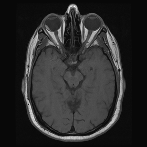
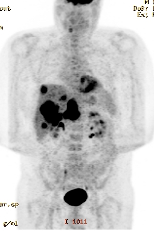
The patient was a 53 year old Caucasian male with a past medical history significant for hypertension. In July of 2009 he visited his care provider with complaints of episodic right eye pain for 9 months with concurrent blind spots. Fundus photography revealed a 9DD (horizontal) by 10DD (vertical) pigmented lesion that was 2mm temporal to the fovea. Fluorescein angiography revealed mottled staining with a rim of block fluorescence extending 1DD temporal to the fovea, and a B-scan showed a temporal choroidal lesion measuring 14.65 x 13.37 x 4.65mm with no extra-scleral extension. An MRI-brain was performed (see below). Further work-up was negative for metastatic disease, and an intensive course of treatment was pursued. However, the patient returned in May 2010 with abdominal pain concerning for metastatic disease. An MRI-abdomen was performed followed by liver biopsy. Despite further treatment, the patient's clinical course deteriorated and the patient expired in November 2010. An autopsy was performed.
Imaging:
Gross images:
|
Autopsy - Right eye |
Autopsy - Liver in situ |
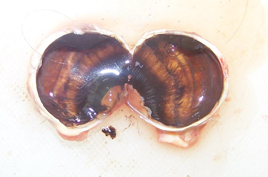 |
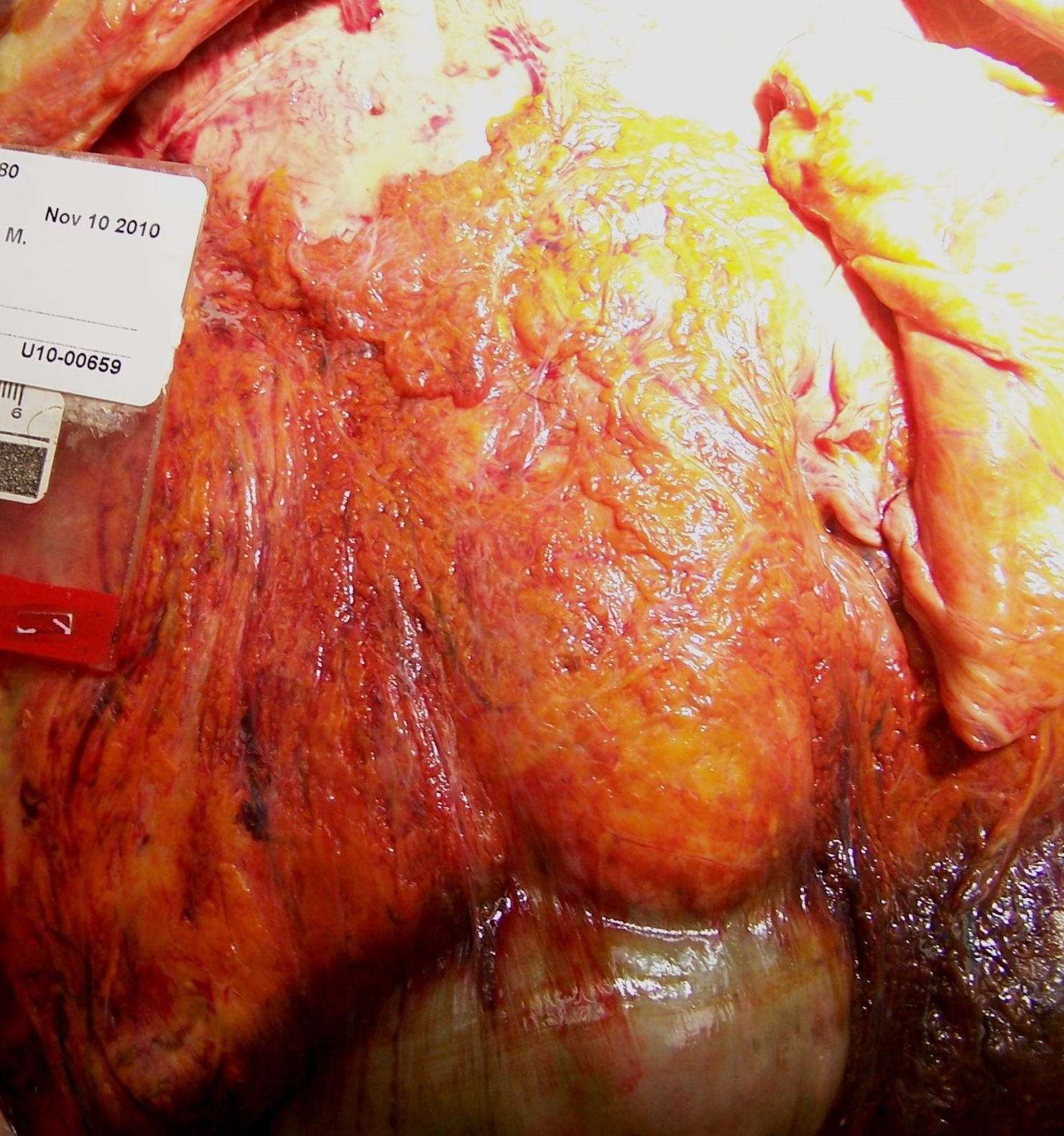 |
Microscopic photographs:
|
Right eye, H&E low power |
Right eye, H&E high power |
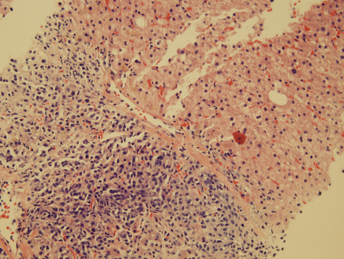
Liver core biopsy, H&E |
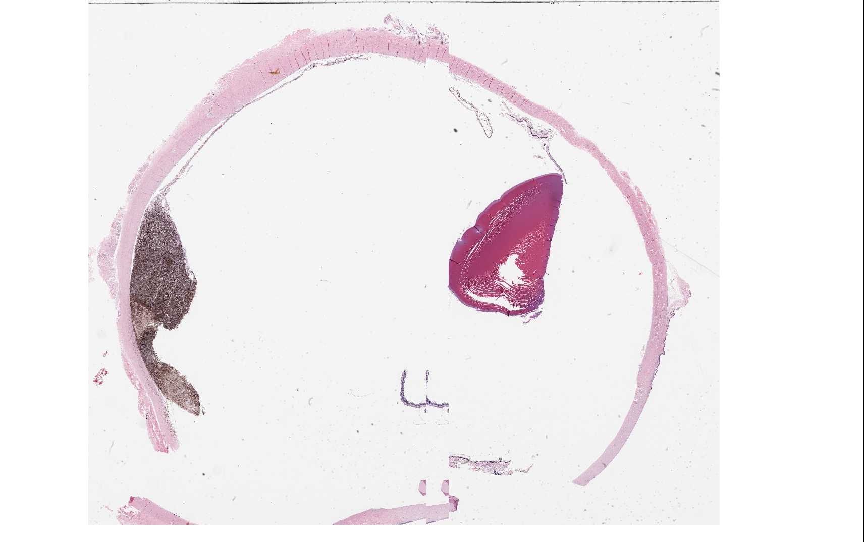 |
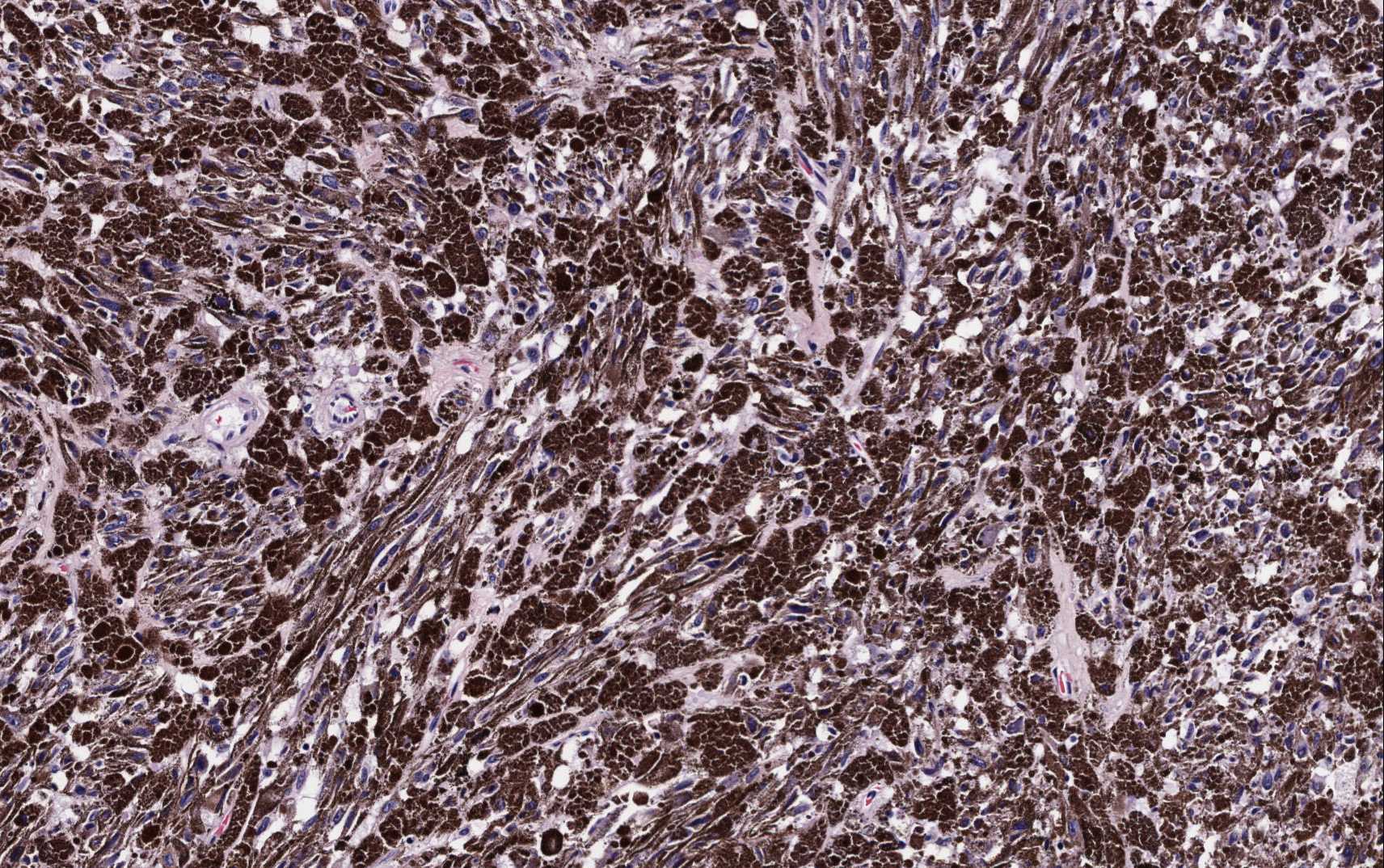 |
 |
|
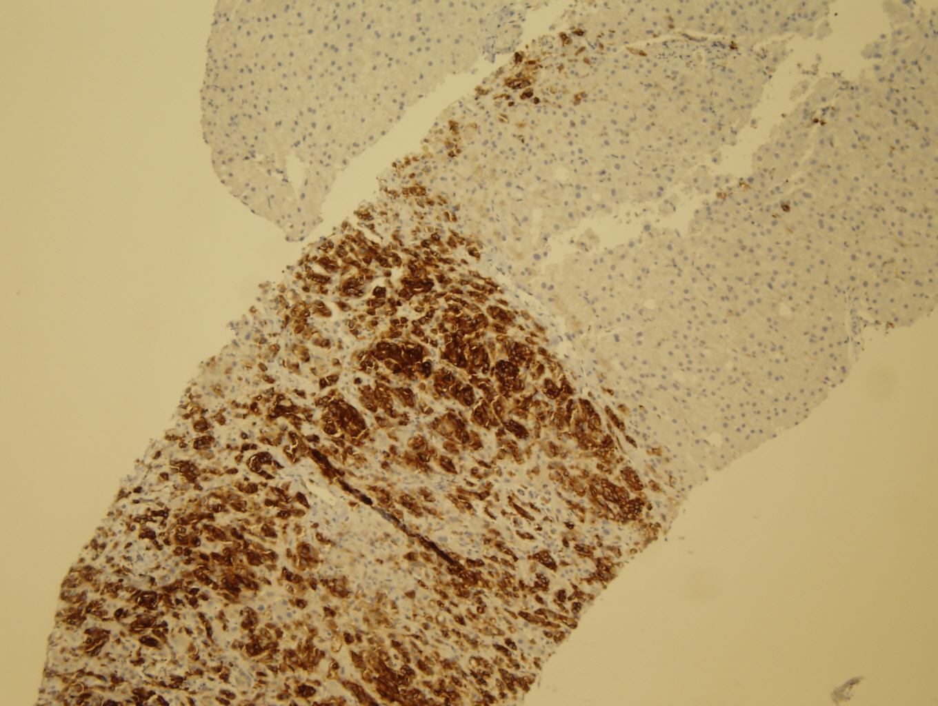
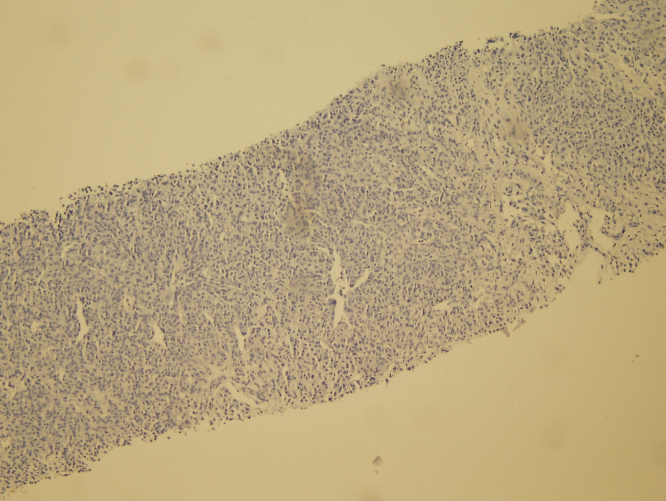
Liver core biopsy, CD117 |
Liver core biopsy, S100 |
Liver core biopsy, HMB45 |
 |
 |
 |


 Meet our Residency Program Director
 LeShelle May
 Chancellor Gary May