
Residency Program - Case of the Month
October 2010 - Presented by Sarah Barnhard, M.D.
Clinical history:
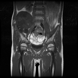
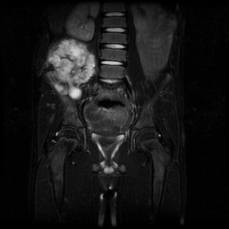
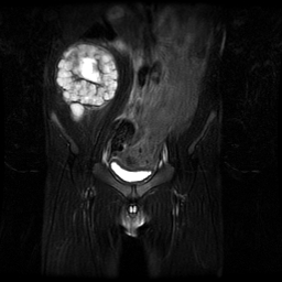
The patient was a 9 year old boy with no past medical history who had a right flank mass which had been growing over several months. MRI of the pelvis revealed a lobulated mass arising from the right iliac wing measuring 10.0cm in greatest dimension. The mass penetrated through the iliac bone, protruded to the posterior aspect of the iliac wing, and displaced the right psoas muscle medially with no radiological evidence of invasion of adjacent structures. Post-gadolinium images revealed heterogeneous enhancement along the margin and septa within the tumor.
 |
 |
 |
|
Gross description:
The specimen consisted of a 353-gram resection of bone and soft tissue measuring in total 8.0cm from medial to lateral x 7.5cm from anterior to posterior x 10.0cm from superior to inferior. A large section of bone was oriented by the surgeon to be the right iliac wing. The remaining tissue was oriented by the surgeon to be one large spherical soft tissue lesion measuring 9.0 x 8.0 x 7.0 cm. Serial sectioning of the tumor revealed variegated surfaces with several areas of cystic degeneration measuring up to 2.0cm in greatest dimension and ranging in color from clear white to pale yellow.
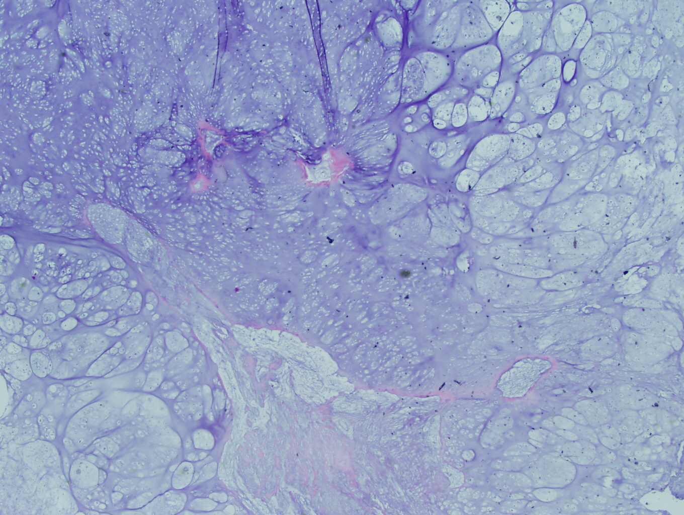
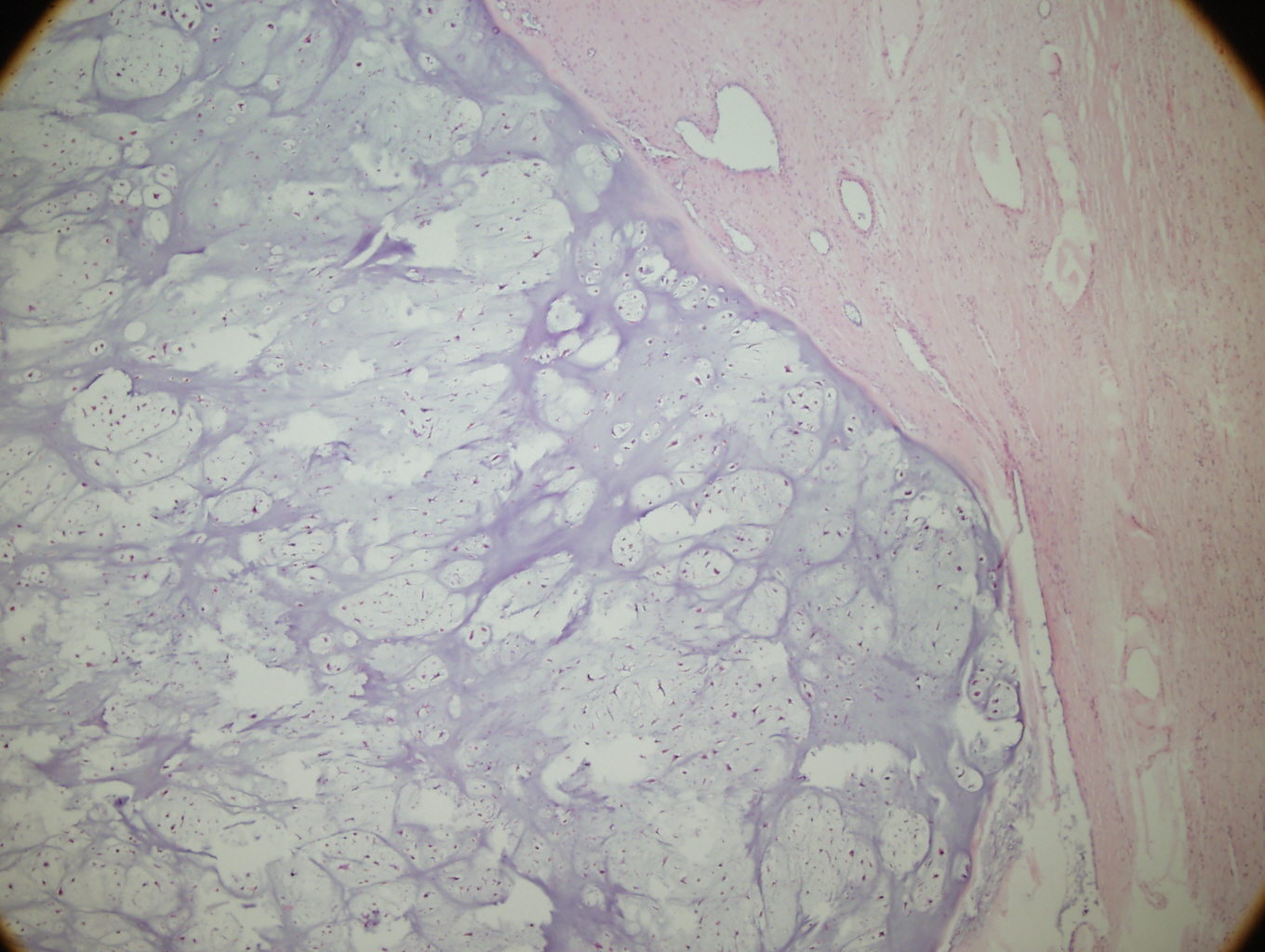
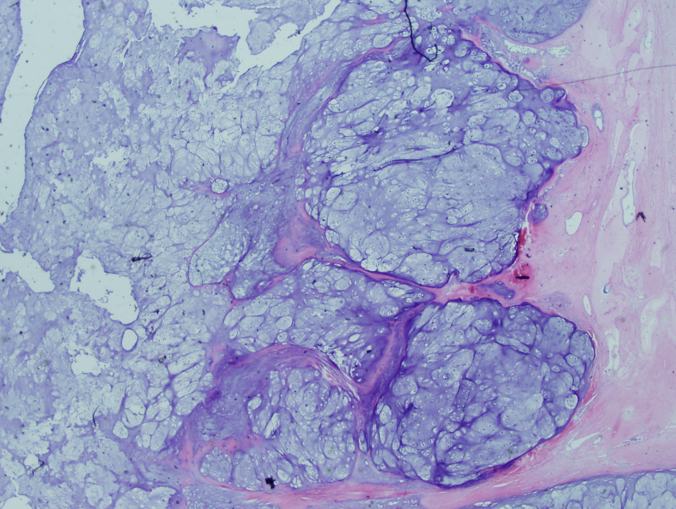
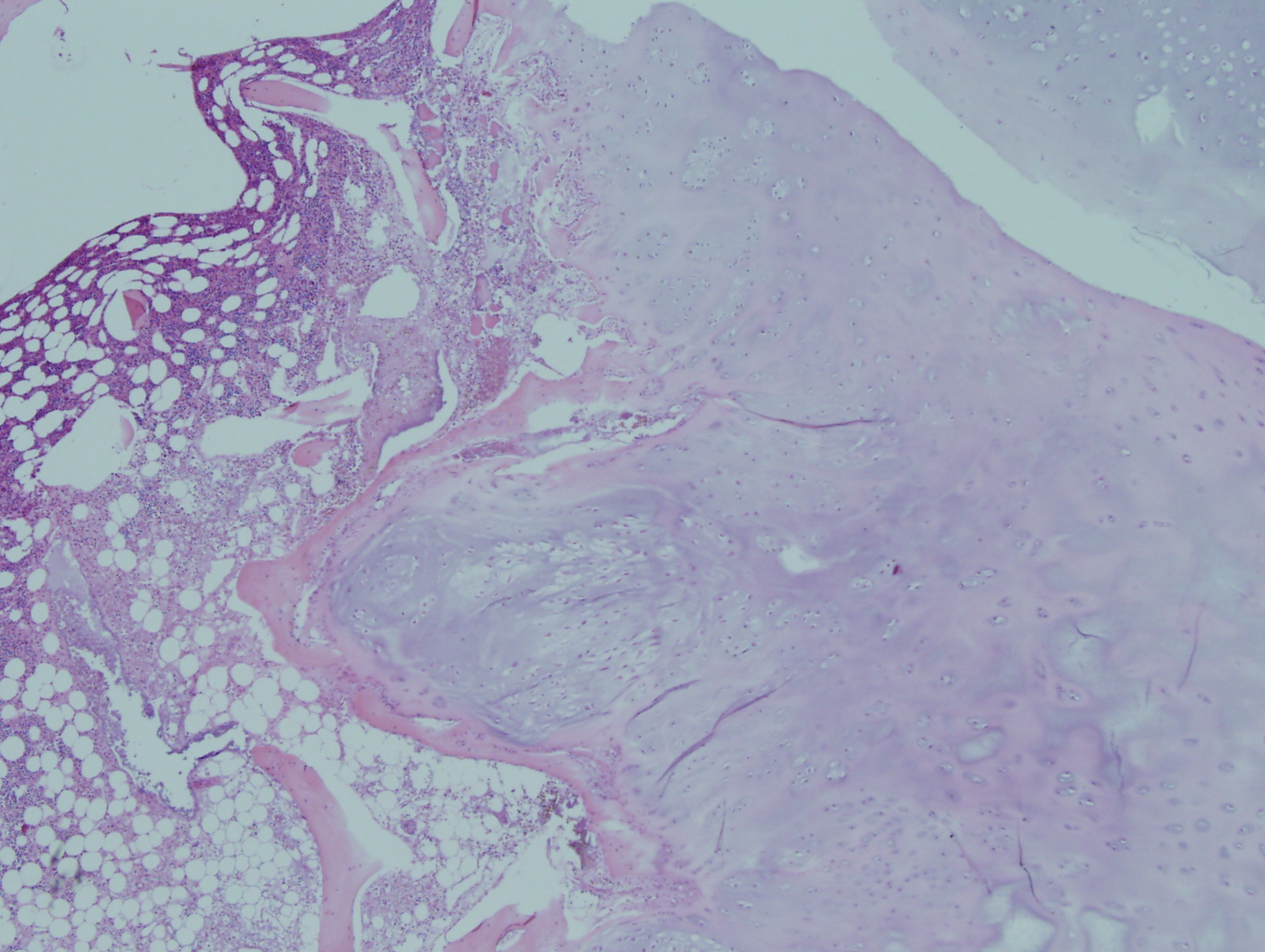
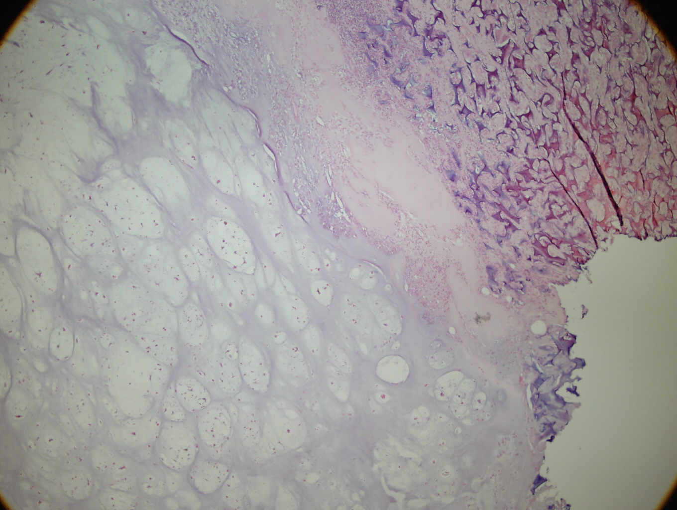
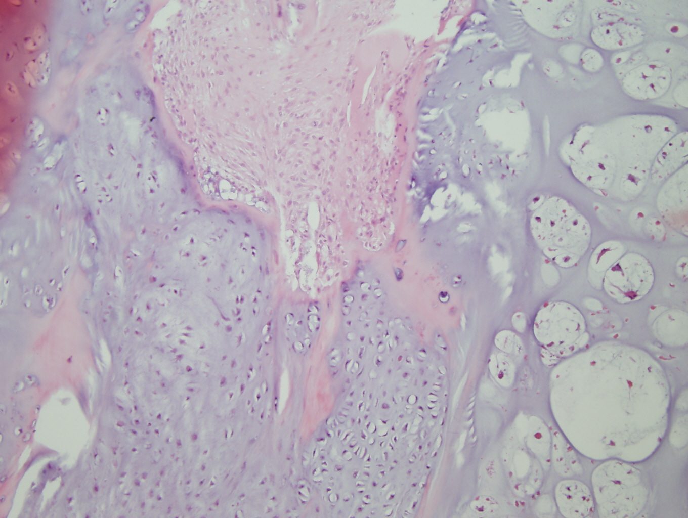
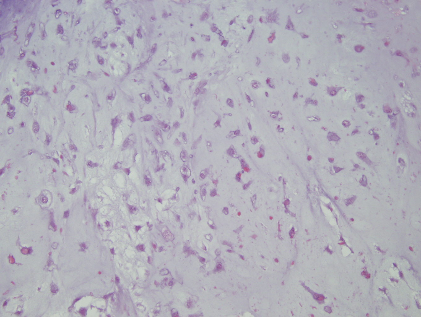
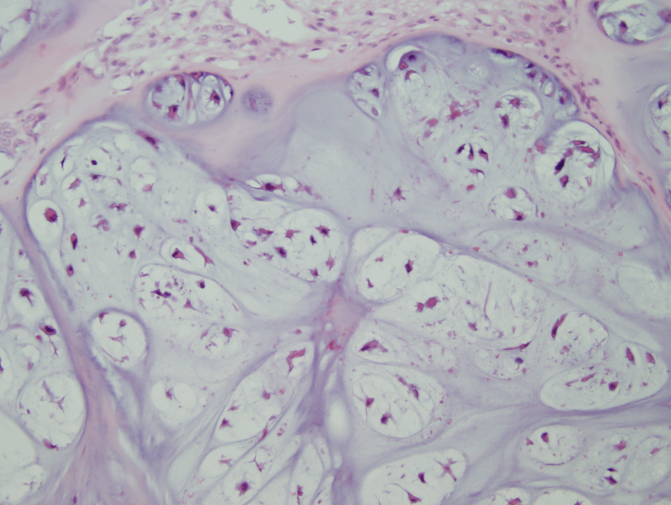
Histology:
The microscopic exam revealed a hyaline and myxoid cartilaginous tumor of low to moderate cellularity as seen in the following pictures:
 Meet our Residency Program Director
 LeShelle May
 Chancellor Gary May