
Residency Program - Case of the Month
September 2010 - Presented by Heidi Jess, M.D.
Clinical history:
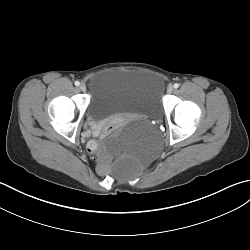
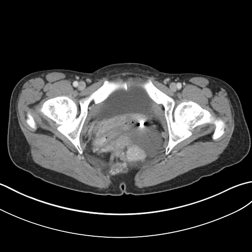
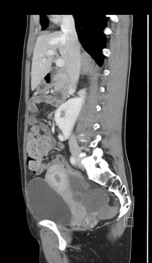
The patient is a 31 year old female with a prior history of a myelomeningocele who was noted to have a new cystic structure within the pelvis/sacrum region at the time of a C-section. Follow-up CT imaging revealed a large, complex cystic mass within the sacrum which extended into the left hemipelvis, measuring 17.0 cm in greatest dimension. She underwent an exploratory laparotomy with subsequent TAH-BSO and resection of soft tissue tumor.
 |
 |
 |
Gross description:
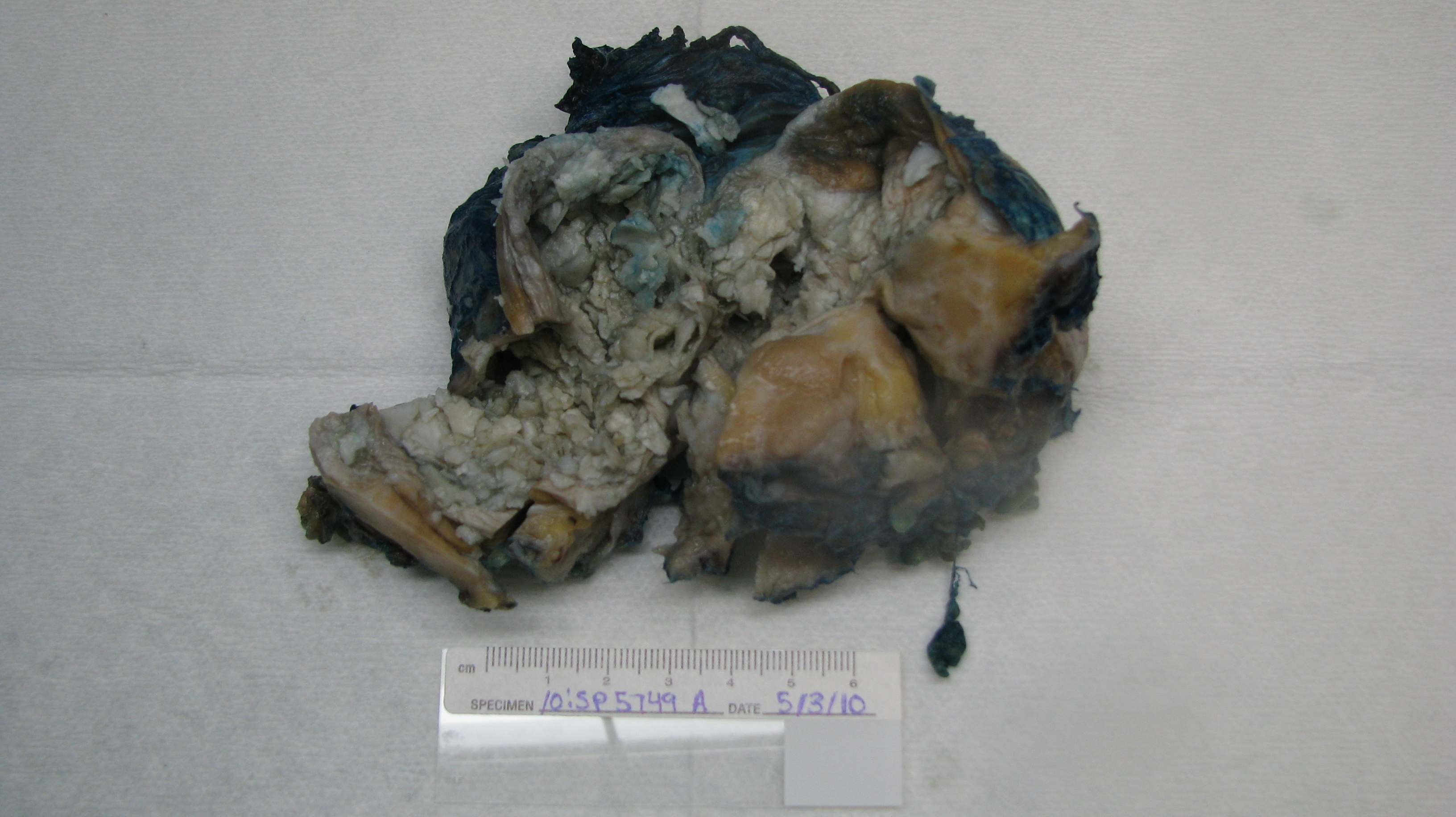
The specimen consisted of a 372 gram composite resection including the cervix, uterus, bilateral ovaries and fallopian tubes and an attached soft tissue mass. The cervix and uterus were unremarkable. A 4.0 cm simple cyst was present within the right ovary, filled with thin chocolate colored fluid. The left ovary contained multiple small simple cysts filled with clear fluid. The soft tissue mass measured 9.0 x 8.0 x 6.0 cm. Serial sections of the mass revealed multiple cystic spaces filled with flaky white-yellow material. Areas of solid tan tissue and yellow adipose tissue were also present.
 |
 |
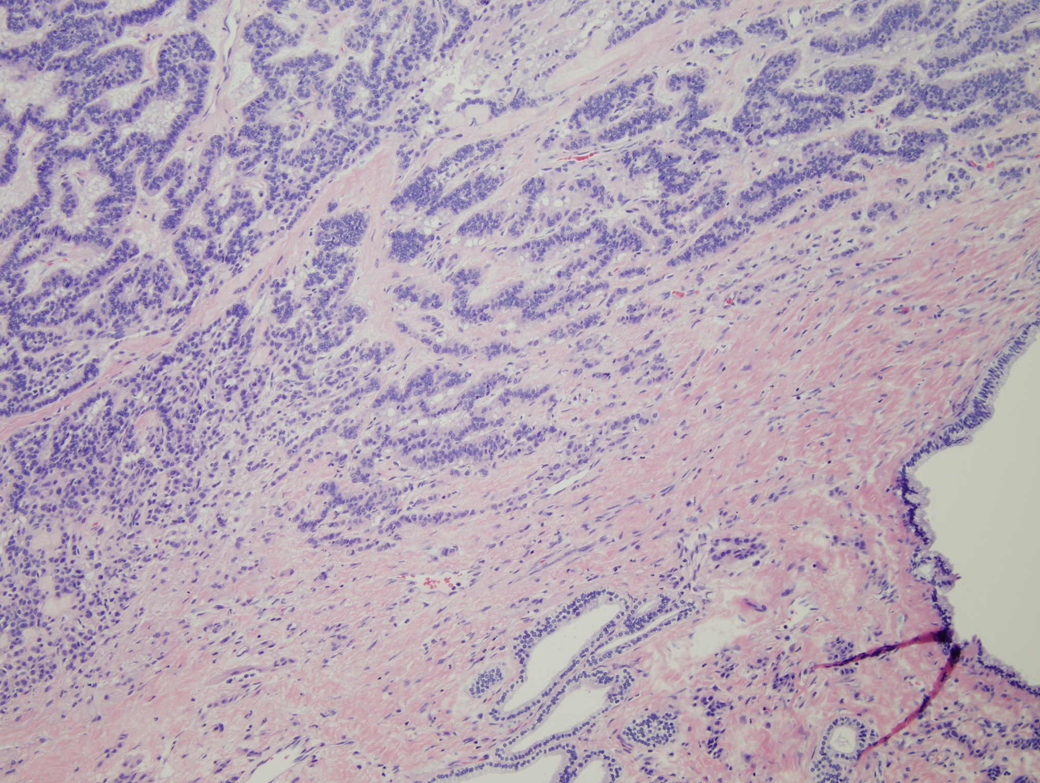
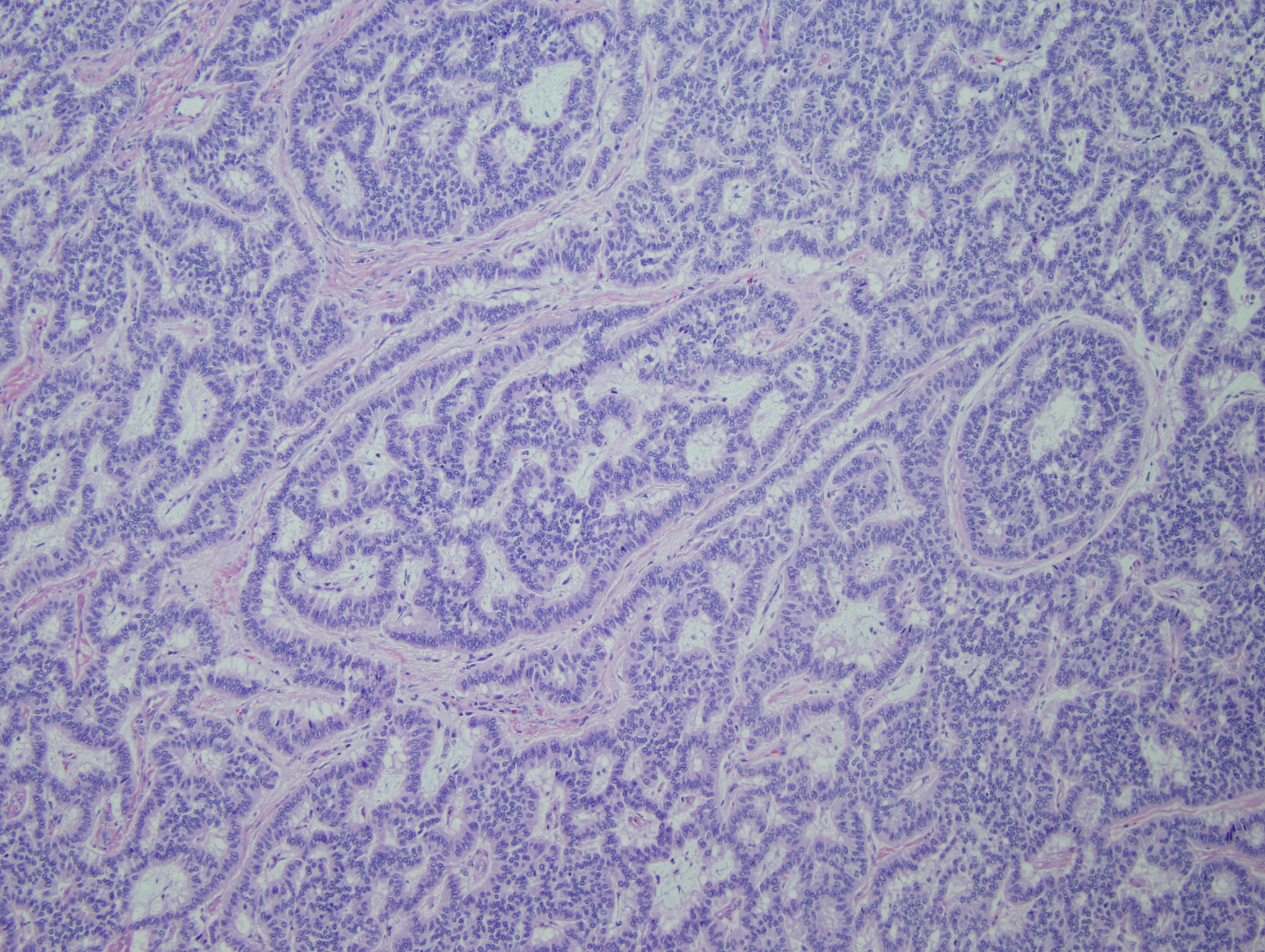
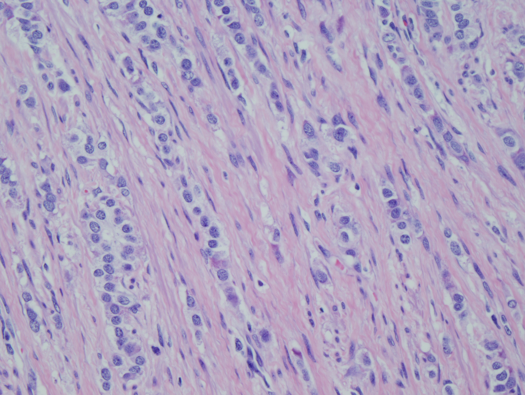
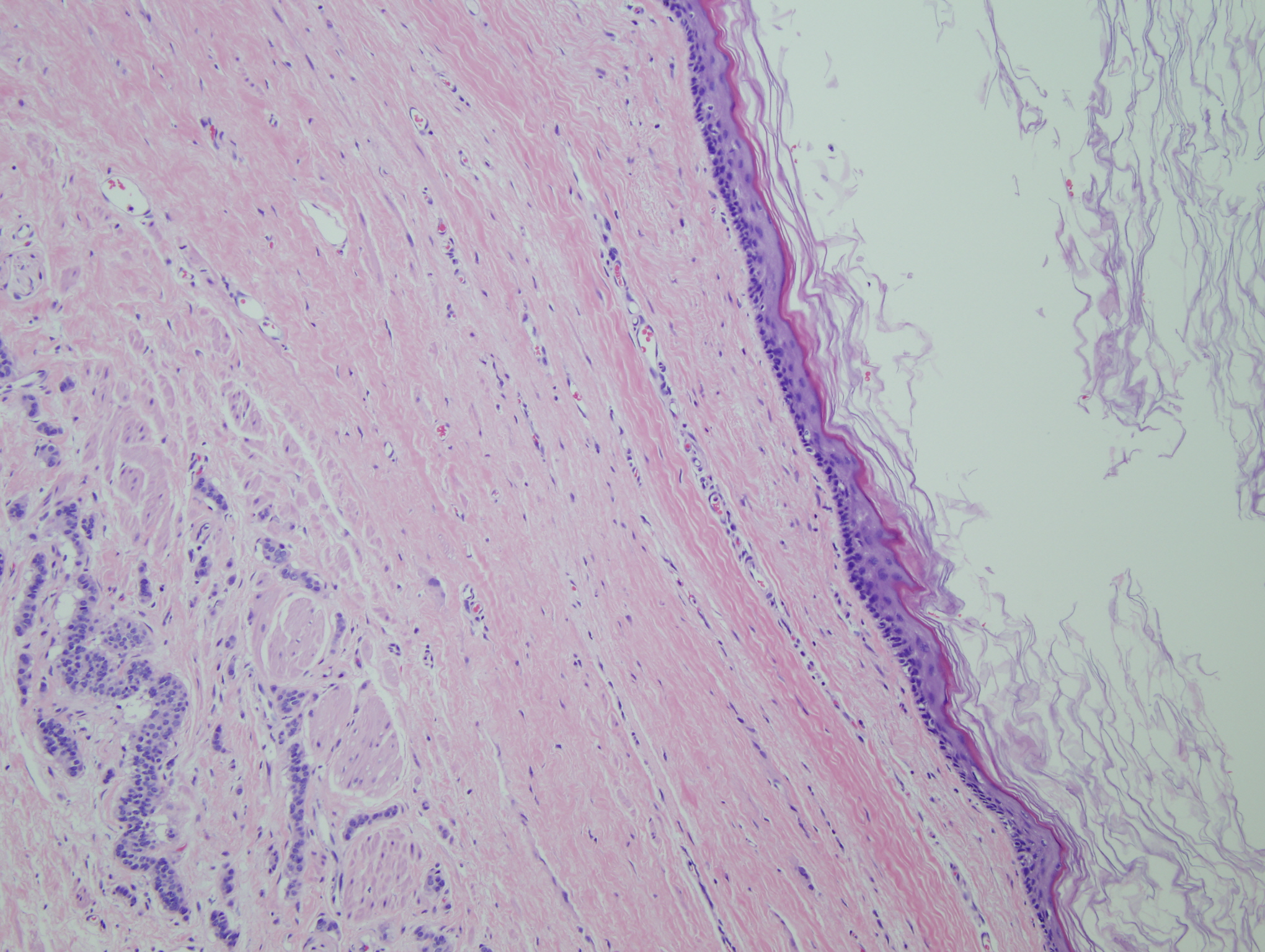
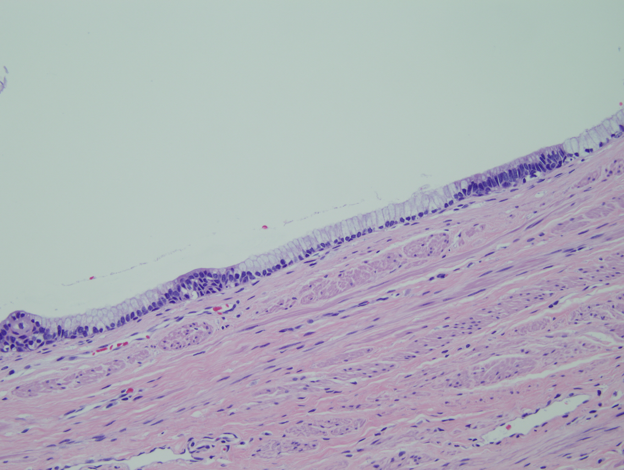
Histology:
Microscopically the tumor was heterogenous with cystic and solid areas as shown in the following figures.

 Meet our Residency Program Director
 LeShelle May
 Chancellor Gary May