MRI Arthrogram
What is an MRI Arthrogram?
MRI arthrogram exam is an imaging study that is used to take detailed pictures of your joints. This exam has 2 parts. First you will have an arthrogram and then MRI. This 2-part exam shows more details of your joint than an MRI by itself. Not every patient will need an arthrogram MRI exam, but in some cases information provided by this type of exam will help your healthcare provider decide which treatment is best for your medical condition.
How do I prepare for my Arthrogram MRI exam?
- You will be asked to arrive 20 minutes prior to your appointment
- During check-in process you will be asked to fill out the MRI Safety From. Click Here to view the form.
- You will be asked to change into a hospital gown and lock up your belongings. Click Here to view safety page.
- You will meet with the doctors before your exam.
- Bring a list of all medicines that you are taking.
- Prepare any questions you might have and ask them at this time.
- You must give written consent before your MRI arthrogram can begin.
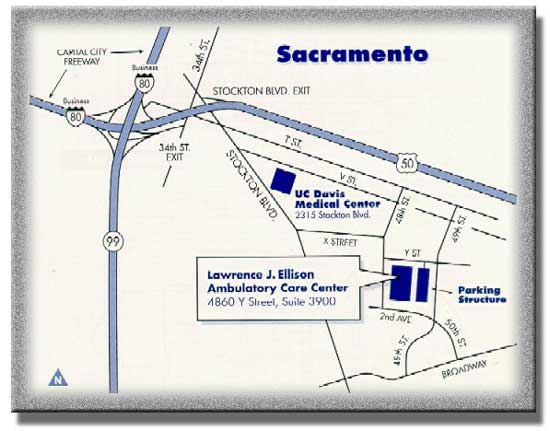
- Arthrogram MRI exams are done only in ACC (Ambulatory Care Center). Click here for directions.
What will happen during my Arthrogram MRI exam?
Part 1- Arthrogram
- Your skin will be cleaned with an antiseptic soap.
- The radiologist will then use a needle to numb the area with a local anesthetic (a numbing medicine).
- When the area is numb, a needle will be placed into your joint space. A fluoroscope will be used to guide the needle.
- When the needle is in the correct place, contrast will be injected. Your joint may feel “full,” or you may feel some pressure in the joint.
- X-ray images will then be taken.
- The arthrogram will take about 30 minutes. Then, you will be sent to the MRI room for part 2 of the study.
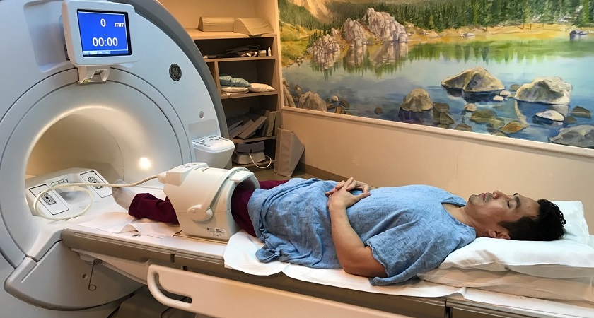
Part 2 - MRI
- You will lie on a sliding table. The MRI technologist will help you get into position for the exam.
- A device called a surface coil will be placed around the joint that is being examined.
- The MRI technologist will position you comfortably in the MRI scanner and then step out of the room to take the images.
- You will be asked not to move while the pictures are being taken.
- Your MRI may take up to 45 minutes.
-
After the exam ends, the doctors in the radiology department will look at your images and provide a report that will be available to your referring doctor, typically within 1-2 days.
Musculoskeletal MRI
How do I prepare for my Musculoskeletal MRI exam?
- You will be asked to arrive 20 minutes prior to your appointment.
- During check-in process you will be asked to fill out the MRI Safety Form, Click Here to view the form.
- You will be asked to change into a hospital gown and lock up your belongings.
- Unless your exam will be done under General Anesthesia, there is no preparation and you can eat and drink prior to your exam.
- Musculoskeletal MRI exams are done in all locations, for directions please click here for directions.
What will happen during my Musculoskeletal MRI exam?
- During the exam you may be given an injection of contrast fluid called gadolinium. This will be administered through a very small IV line in your arm.
- Your MRI can last anywhere from 30 minutes to more than an hour.
- You will be given earplugs as the magnet creates a knocking sound during the exam. If you are claustrophobic, you may want to ask the doctor who ordered this MRI for a mild sedative to take before the exam. Please click here to be directed to our Comfort page.
- You must hold very still because movement can blur the resulting images and delay the completion of the exam.
- There are no restrictions after your exam and you may eat and drink as usual.
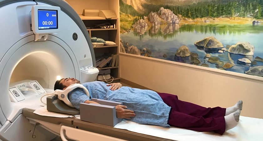
Patient prepared for a Knee MRI
Patient prepared for a Shoulder MRI